2025
Rapti A, Kyrousi C.
Gene Expression Manipulation Via Acute Electroporation in Human Brain Organoids. Methods in molecular biology (Clifton, N.J.) [Internet]. 2025;2899:221-232.
PubmedAbstractBrain organoids are in vitro 3D cultures generated in the lab from human induced pluripotent stem cells or embryonic stem cells and can mimic the human brain structure and function. Specifically, they reproduce to some extent in vivo developmental events as they consist of diverse cell types, such as apical radial glial cells, intermediate progenitors, basal radial glial cells, and neurons forming stratified cortical layers similar to what is observed in the human brain in vivo. Due to cytoarchitecture similarities between the human brain and brain organoids, the latter have been proposed as excellent models for studying human brain development and disease. Thus, genome manipulation in brain organoids is crucial for investigating the functions of specific genes and mutations that have been associated with brain-related disorders. For this reason, gene manipulation has been implemented in brain organoids in the last few years. Here, we describe a step-by-step protocol for gene expression manipulation and analyses in brain organoids via acute electroporation that we have optimized based on the in vivo electroporation that has been widely used in animal models. This easy-to-apply protocol is fast and robust and facilitates the precise spatiotemporal manipulation of the expression of any gene of interest.
2023
Pipicelli F, Baumann N, Di Giaimo R, Forero-Echeverry A, Kyrousi C, Bonrath R, Maccarrone G, Jabaudon D, Cappello S.
Non–cell-autonomous regulation of interneuron specification mediated by extracellular vesicles. Science Advances [Internet]. 2023;9(20):eadd8164.
Pubmed AbstractDisruption in neurogenesis and neuronal migration can influence the assembly of cortical circuits, affecting the excitatory-inhibitory balance and resulting in neurodevelopmental and neuropsychiatric disorders. Using ventral cerebral organoids and dorsoventral cerebral assembloids with mutations in the extracellular matrix gene LGALS3BP, we show that extracellular vesicles released into the extracellular environment regulate the molecular differentiation of neurons, resulting in alterations in migratory dynamics. To investigate how extracellular vesicles affect neuronal specification and migration dynamics, we collected extracellular vesicles from ventral cerebral organoids carrying a mutation in LGALS3BP, previously identified in individuals with cortical malformations and neuropsychiatric disorders. These results revealed differences in protein composition and changes in dorsoventral patterning. Proteins associated with cell fate decision, neuronal migration, and extracellular matrix composition were altered in mutant extracellular vesicles. Moreover, we show that treatment with extracellular vesicles changes the transcriptomic profile in neural progenitor cells. Our results indicate that neuronal molecular differentiation can be influenced by extracellular vesicles.
2022
Cruceanu C, Dony L, Krontira AC, Fischer DS, Roeh S, Di Giaimo R, Kyrousi C, Kaspar L, Arloth J, Czamara D.
Cell-type-specific impact of glucocorticoid receptor activation on the developing brain: A Cerebral organoid study. American Journal of Psychiatry [Internet]. 2022;179(5):375-387.
Pubmed Abstract
Objective: A fine-tuned balance of glucocorticoid receptor (GR) activation is essential for organ formation, with disturbances influencing many health outcomes. In utero, glucocorticoids have been linked to brain-related negative outcomes, with unclear underlying mechanisms, especially regarding cell-type-specific effects. An in vitro model of fetal human brain development, induced human pluripotent stem cell (hiPSC)-derived cerebral organoids, was used to test whether cerebral organoids are suitable for studying the impact of prenatal glucocorticoid exposure on the developing brain.
Methods: The GR was activated with the synthetic glucocorticoid dexamethasone, and the effects were mapped using single-cell transcriptomics across development.
Results: The GR was expressed in all cell types, with increasing expression levels through development. Not only did its activation elicit translocation to the nucleus and the expected effects on known GR-regulated pathways, but also neurons and progenitor cells showed targeted regulation of differentiation- and maturation-related transcripts. Uniquely in neurons, differentially expressed transcripts were significantly enriched for genes associated with behavior-related phenotypes and disorders. This human neuronal glucocorticoid response profile was validated across organoids from three independent hiPSC lines reprogrammed from different source tissues from both male and female donors.
Conclusions: These findings suggest that excessive glucocorticoid exposure could interfere with neuronal maturation in utero, leading to increased disease susceptibility through neurodevelopmental processes at the interface of genetic susceptibility and environmental exposure. Cerebral organoids are a valuable translational resource for exploring the effects of glucocorticoids on early human brain development.
Keywords: Biology; Brain; Child/Adolescent Psychiatry; Development; Glucocorticoid Receptor; Neurodevelopmental Disorders; Pre/Peri/Postnatal Issues; Stress; Translational Research.
Angelopoulos I, Gakis G, Birmpas K, Kyrousi C, Habeos EI, Kaplani K, Lygerou Z, Habeos I, Taraviras S.
Metabolic regulation of the neural stem cell fate: unravelling new connections, establishing new concepts. Frontiers in Neuroscience [Internet]. 2022:1800.
Pubmed Abstract
The neural stem cell niche is a key regulator participating in the maintenance, regeneration, and repair of the brain. Within the niche neural stem cells (NSC) generate new neurons throughout life, which is important for tissue homeostasis and brain function. NSCs are regulated by intrinsic and extrinsic factors with cellular metabolism being lately recognized as one of the most important ones, with evidence suggesting that it may serve as a common signal integrator to ensure mammalian brain homeostasis. The aim of this review is to summarize recent insights into how metabolism affects NSC fate decisions in adult neural stem cell niches, with occasional referencing of embryonic neural stem cells when it is deemed necessary. Specifically, we will highlight the implication of mitochondria as crucial regulators of NSC fate decisions and the relationship between metabolism and ependymal cells. The link between primary cilia dysfunction in the region of hypothalamus and metabolic diseases will be examined as well. Lastly, the involvement of metabolic pathways in ependymal cell ciliogenesis and physiology regulation will be discussed.
Keywords: cell mechanics; ciliopathies; ependymal; metabolism; neural stem cell niche; neural stem cells; subventricular zone (SVZ).
Damianidou E*, Mouratidou L*, Kyrousi C.
Research models of neurodevelopmental disorders: The right model in the right place. *Equal contributing authors. Frontiers in Neuroscience [Internet]. 2022:1846.
Pubmed Abstract
Neurodevelopmental disorders (NDDs) are a heterogeneous group of impairments that affect the development of the central nervous system leading to abnormal brain function. NDDs affect a great percentage of the population worldwide, imposing a high societal and economic burden and thus, interest in this field has widely grown in recent years. Nevertheless, the complexity of human brain development and function as well as the limitations regarding human tissue usage make their modeling challenging. Animal models play a central role in the investigation of the implicated molecular and cellular mechanisms, however many of them display key differences regarding human phenotype and in many cases, they partially or completely fail to recapitulate them. Although in vitro two-dimensional (2D) human-specific models have been highly used to address some of these limitations, they lack crucial features such as complexity and heterogeneity. In this review, we will discuss the advantages, limitations and future applications of in vivo and in vitro models that are used today to model NDDs. Additionally, we will describe the recent development of 3-dimensional brain (3D) organoids which offer a promising approach as human-specific in vitro models to decipher these complex disorders.
Kyrousi C*, Taraviras S*.
The role of brain organoids as model system for human disease. *Co-corresponding authors. Editorial in Achaiki Iatriki [Internet]. 2022:41(2): 69–72.
Achaiki Iatriki Krontira AC, Cruceanu C, Kyrousi C, Dony L, Link M-H, Kappelmann N, Pöhlchen D, Roeh S, Sportelli V, Wölfel B.
Temporal regulation of ZBTB16 expression by glucocorticoids alters human cortical neurogenesis. bioRxiv [Internet]. 2022:2022.08. 21.504700.
bioRxiv AbstractGlucocorticoids are important for proper organ maturation
1. Increased exposure to these hormones during pregnancy, as a result of commonly prescribed synthetic glucocorticoids such as dexamethasone in preterm births
2, has been associated with lasting effects on the offspring, including on neurodevelopment and neuropsychiatric disease risk
3. While the consequences of glucocorticoid excess in term and especially adult brain have been extensively studied, mainly in rodents
4, studies on their effects during early human cortical development are absent. Here we use human cerebral organoids and mice to study cell-type specific effects of glucocorticoids on neurogenic processes. We show that glucocorticoid administration during neurogenesis alters the cellular architecture of the developing cortex by increasing a specific type of gyrencephalic species-enriched basal progenitors that co-express
PAX6 and
EOMES. This effect is mediated via the glucocorticoid-responsive transcription factor
ZBTB16 as shown with overexpression, genetic knock-down and reporter assays experiments in organoids and embryonic mouse models and leads to increased production of deep-layer neurons. A phenome-wide mendelian randomization analysis of a genetic intronic enhancer variant that moderates glucocorticoid-induced
ZBTB16 levels, as shown with enhancer assays and enhancer-editing in organoids, reveals potential causal relationships with increased educational attainment as well as neuroimaging phenotypes in adults. In this study we provide a cellular and molecular pathway for the effects of glucocorticoids on human neurogenesis that potentially explains postnatal phenotypes and may be used to refine treatment guidelines.
2021
Kyrousi C, O’Neill AC, Brazovskaja A, He Z, Kielkowski P, Coquand L, Di Giaimo R, D’Andrea P, Belka A, Forero Echeverry A.
Extracellular LGALS3BP regulates neural progenitor position and relates to human cortical complexity. Nature Communications [Internet]. 2021;12(1):6298.
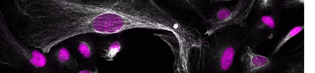
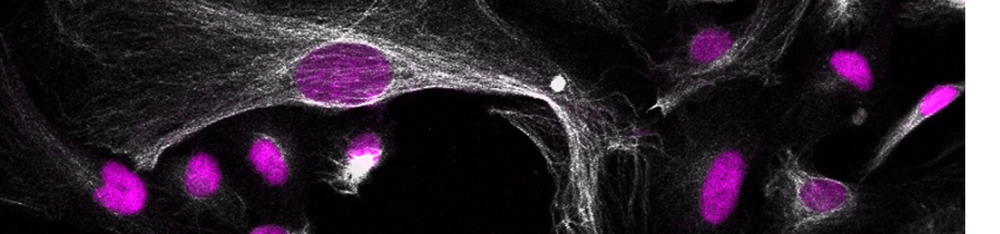
Pubmed AbstractBasal progenitors (BPs), including intermediate progenitors and basal radial glia, are generated from apical radial glia and are enriched in gyrencephalic species like humans, contributing to neuronal expansion. Shortly after generation, BPs delaminate towards the subventricular zone, where they further proliferate before differentiation. Gene expression alterations involved in BP delamination and function in humans are poorly understood. Here, we study the role of LGALS3BP, so far known as a cancer biomarker, which is a secreted protein enriched in human neural progenitors (NPCs). We show that individuals with LGALS3BP de novo variants exhibit altered local gyrification, sulcal depth, surface area and thickness in their cortex. Additionally, using cerebral organoids, human fetal tissues and mice, we show that LGALS3BP regulates the position of NPCs. Single-cell RNA-sequencing and proteomics reveal that LGALS3BP-mediated mechanisms involve the extracellular matrix in NPCs' anchoring and migration within the human brain. We propose that its temporal expression influences NPCs' delamination, corticogenesis and gyrification extrinsically.
2020
Di Matteo F*, Pipicelli F*, Kyrousi C, Tovecci I, Penna E, Crispino M, Chambery A, Russo R, Ayo‐Martin AC, Giordano M, et al. Cystatin B is essential for proliferation and interneuron migration in individuals with EPM 1 epilepsy. * Equal contributing authors. EMBO molecular medicine [Internet]. 2020;12(6):e11419.
Pubmed AbstractProgressive myoclonus epilepsy (PME) of Unverricht-Lundborg type (EPM1) is an autosomal recessive neurodegenerative disorder with the highest incidence of PME worldwide. Mutations in the gene encoding cystatin B (CSTB) are the primary genetic cause of EPM1. Here, we investigate the role of CSTB during neurogenesis in vivo in the developing mouse brain and in vitro in human cerebral organoids (hCOs) derived from EPM1 patients. We find that CSTB (but not one of its pathological variants) is secreted into the mouse cerebral spinal fluid and the conditioned media from hCOs. In embryonic mouse brain, we find that functional CSTB influences progenitors' proliferation and modulates neuronal distribution by attracting interneurons to the site of secretion via cell-non-autonomous mechanisms. Similarly, in patient-derived hCOs, low levels of functional CSTB result in an alteration of progenitor's proliferation, premature differentiation, and changes in interneurons migration. Secretion and extracellular matrix organization are the biological processes particularly affected as suggested by a proteomic analysis in patients' hCOs. Overall, our study sheds new light on the cellular mechanisms underlying the development of EPM1.
Buchsbaum IY, Kielkowski P, Giorgio G, O'Neill AC, Di Giaimo R, Kyrousi C, Khattak S, Sieber SA, Robertson SP, Cappello S.
ECE 2 regulates neurogenesis and neuronal migration during human cortical development. EMBO reports [Internet]. 2020;21(5):e48204.
Pubmed AbstractDuring embryonic development, excitatory projection neurons migrate in the cerebral cortex giving rise to organised layers. Periventricular heterotopia (PH) is a group of aetiologically heterogeneous disorders in which a subpopulation of newborn projection neurons fails to initiate their radial migration to the cortex, ultimately resulting in bands or nodules of grey matter lining the lateral ventricles. Although a number of genes have been implicated in its cause, currently they only satisfactorily explain the pathogenesis of the condition for 50% of patients. Novel gene discovery is complicated by the extreme genetic heterogeneity recently described to underlie its cause. Here, we study the neurodevelopmental role of endothelin-converting enzyme-2 (ECE2) for which two biallelic variants have been identified in two separate patients with PH. Our results show that manipulation of ECE2 levels in human cerebral organoids and in the developing mouse cortex leads to ectopic localisation of neural progenitors and neurons. We uncover the role of ECE2 in neurogenesis, and mechanistically, we identify its involvement in the generation and secretion of extracellular matrix proteins in addition to cytoskeleton and adhesion.
Ayo-Martin AC, Kyrousi C, Di Giaimo R, Cappello S.
GNG5 controls the number of apical and basal progenitors and alters neuronal migration during cortical development. Frontiers in Molecular Biosciences [Internet]. 2020;7:578137.
Pubmed AbstractCortical development is a very complex process in which any temporal or spatial alterations can give rise to a wide range of cortical malformations. Among those malformations, periventricular heterotopia (PH) is characterized by clusters of neurons that do not migrate to the correct place. Cerebral organoids derived from patients with mutations in DCHS1 and FAT4, which have been associated with PH, exhibit higher levels of GNG5 expression in a patient-specific cluster of neurons. Here we investigate the role of GNG5 during the development of the cerebral cortex in mice and human cerebral organoids. GNG5, highly expressed in progenitors and downregulated in neurons, is critical for controlling the number of apical and basal progenitors and neuronal migration. Moreover, forced expression of GNG5 recapitulates some of the alterations observed upon downregulation of Dchs1 and Fat4 in mice and human cerebral organoids derived from DCHS1 and FAT4 patients, suggesting a critical role of GNG5 in cortical development.
Kullmann JA*, Meyer S*, Pipicelli F*, Kyrousi C, Schneider F, Bartels N, Cappello S, Rust MB.
Profilin1-dependent F-actin assembly controls division of apical radial glia and neocortex development. *Equal contributing authors. Cerebral Cortex [Internet]. 2020;30(6):3467-3482.
Pubmed AbstractNeocortex development depends on neural stem cell proliferation, cell differentiation, neurogenesis, and neuronal migration. Cytoskeletal regulation is critical for all these processes, but the underlying mechanisms are only poorly understood. We previously implicated the cytoskeletal regulator profilin1 in cerebellar granule neuron migration. Since we found profilin1 expressed throughout mouse neocortex development, we here tested the hypothesis that profilin1 is crucial for neocortex development. We found no evidence for impaired neuron migration or layering in the neocortex of profilin1 mutant mice. However, proliferative activity at basal positions was doubled in the mutant neocortex during mid-neurogenesis, with a drastic and specific increase in basal Pax6+ cells indicative for elevated numbers of basal radial glia (bRG). This was accompanied by transiently increased neurogenesis and associated with mild invaginations resembling rudimentary neocortex folds. Our data are in line with a model in which profilin1-dependent actin assembly controls division of apical radial glia (aRG) and thereby the fate of their progenies. Via this mechanism, profilin1 restricts cell delamination from the ventricular surface and, hence, bRG production and thereby controls neocortex development in mice. Our data support the radial cone hypothesis" claiming that elevated bRG number causes neocortex folds.
Tsaridou S, Skamnelou M, Iliadou M, Lokka G, Parlapani E, Mougkogianni M, Danalatos R-I, Kanellou A, Chlorogiannis D-D, Kyrousi C, et al. Three-dimensional models for studying neurodegenerative and neurodevelopmental diseases. GeNeDis 2018: Genetics and Neurodegeneration [Internet]. 2020:35-41.
Pubmed AbstractHuman brain possesses a unique anatomy and physiology. For centuries, methodological barriers and ethical challenges in accessing human brain tissues have restricted researchers into using 2-D cell culture systems and model organisms as a tool for investigating the mechanisms underlying neurological disorders in humans. However, our understanding regarding the human brain development and diseases has been recently extended due to the generation of 3D brain organoids, grown from human stem cells or induced pluripotent stem cells (iPSCs). This system evolved into an attractive model of brain diseases as it recapitulates to a great extend the cellular organization and the microenvironment of a human brain. This chapter focuses on the application of brain organoids in modelling several neurodevelopmental and neurodegenerative diseases.
Drechsel J, Kyrousi C, Cappello S, Sieber SA.
Tranylcypromine specificity for monoamine oxidase is limited by promiscuous protein labelling and lysosomal trapping. RSC Chemical Biology [Internet]. 2020;1(4):209-213.
Pubmed AbstractMonoamine oxidases MAOA and MAOB catalyze important cellular functions such as the deamination of neurotransmitters. Correspondingly, MAO inhibitors are used for the treatment of severe neuropsychiatric disorders such as depression. A commonly prescribed drug against refractory depression is tranylcypromine, however, the side effects are poorly understood. In order to decipher putative off-targets, we synthesized two tranylcypromine probes equipped with either an alkyne moiety or an alkyne-diazirine minimal photocrosslinker for in situ proteome profiling. Surprisingly, LC-MS/MS analysis revealed low enrichment of MAOA and relatively promiscuous labeling of proteins. Photoprobe labeling paired with fluorescent imaging studies revealed lysosomal trapping which could be largely reverted by the addition of lysosomotropic drugs.
Kyrousi C, Cappello S.
Using brain organoids to study human neurodevelopment, evolution and disease. Wiley Interdisciplinary Reviews: Developmental Biology [Internet]. 2020;9(1):e347.
Pubmed AbstractThe brain is one of the most complex organs, responsible for the advanced intellectual and cognitive ability of humans. Although primates are to some extent capable of performing cognitive tasks, their abilities are less evolved. One of the reasons for this is the vast differences in the brain of humans compared to other mammals, in terms of shape, size and complexity. Such differences make the study of human brain development fascinating. Interestingly, the cerebral cortex is by far the most complex brain region resulting from its selective evolution within mammals over millions of years. Unraveling the molecular and cellular mechanisms regulating brain development, as well as the evolutionary differences seen across species and the need to understand human brain disorders, are some of the reasons why scientists are interested in improving their current knowledge on human corticogenesis. Toward this end, several animal models including primates have been used, however, these models are limited in their extent to recapitulate human-specific features. Recent technological achievements in the field of stem cell research, which have enabled the generation of human models of corticogenesis, called brain or cerebral organoids, are of great importance. This review focuses on the main cellular and molecular features of human corticogenesis and the use of brain organoids to study it. We will discuss the key differences between cortical development in human and nonhuman mammals, the technological applications of brain organoids and the different aspects of cortical development in normal and pathological conditions, which can be modeled using brain organoids. This article is categorized under: Comparative Development and Evolution > Regulation of Organ Diversity Nervous System Development > Vertebrates: General Principles.
2019
Lalioti M-E, Arbi M, Loukas I, Kaplani K, Kalogeropoulou A, Lokka G, Kyrousi C, Mizi A, Georgomanolis T, Josipovic N, et al. GemC1 governs multiciliogenesis through direct interaction with and transcriptional regulation of p73. Journal of Cell Science [Internet]. 2019;132(11):jcs228684.
Pubmed AbstractA distinct combination of transcription factors elicits the acquisition of a specific fate and the initiation of a differentiation program. Multiciliated cells (MCCs) are a specialized type of epithelial cells that possess dozens of motile cilia on their apical surface. Defects in cilia function have been associated with ciliopathies that affect many organs, including brain and airway epithelium. Here we show that the geminin coiled-coil domain-containing protein 1 GemC1 (also known as Lynkeas) regulates the transcriptional activation of p73, a transcription factor central to multiciliogenesis. Moreover, we show that GemC1 acts in a trimeric complex with transcription factor E2F5 and tumor protein p73 (officially known as TP73), and that this complex is important for the activation of the p73 promoter. We also provide in vivo evidence that GemC1 is necessary for p73 expression in different multiciliated epithelia. We further show that GemC1 regulates multiciliogenesis through the control of chromatin organization, and the epigenetic marks/tags of p73 and Foxj1. Our results highlight novel signaling cues involved in the commitment program of MCCs across species and tissues.This article has an associated First Person interview with the first author of the paper.
Lalioti M‐E, Kaplani K, Lokka G, Georgomanolis T, Kyrousi C, Dong W, Dunbar A, Parlapani E, Damianidou E, Spassky N, et al. GemC1 is a critical switch for neural stem cell generation in the postnatal brain. Glia [Internet]. 2019;67(12):2360-2373.
Pubmed AbstractThe subventricular zone (SVZ) is one of two main niches where neurogenesis persists during adulthood, as it retains neural stem cells (NSCs) with self-renewal capacity and multi-lineage potency. Another critical cellular component of the niche is the population of postmitotic multiciliated ependymal cells. Both cell types are derived from radial glial cells that become specified to each lineage during embryogenesis. We show here that GemC1, encoding Geminin coiled-coil domain-containing protein 1, is associated with congenital hydrocephalus in humans and mice. Our results show that GemC1 deficiency drives cells toward a NSC phenotype, at the expense of multiciliated ependymal cell generation. The increased number of NSCs is accompanied by increased levels of proliferation and neurogenesis in the postnatal SVZ. Finally, GemC1-knockout cells display altered chromatin organization at multiple loci, further supporting a NSC identity. Together, these findings suggest that GemC1 regulates the balance between NSC generation and ependymal cell differentiation, with implications for the pathogenesis of human congenital hydrocephalus.
Klaus J*, Kanton S*, Kyrousi C*, Ayo-Martin AC, Di Giaimo R, Riesenberg S, O’Neill AC, Camp GJ, Tocco C, Santel M, et al. Altered neuronal migratory trajectories in human cerebral organoids derived from individuals with neuronal heterotopia. *Equal contributing authors. Nature medicine [Internet]. 2019;25(4):561-568.
Pubmed AbstractMalformations of the human cortex represent a major cause of disability1. Mouse models with mutations in known causal genes only partially recapitulate the phenotypes and are therefore not unlimitedly suited for understanding the molecular and cellular mechanisms responsible for these conditions2. Here we study periventricular heterotopia (PH) by analyzing cerebral organoids derived from induced pluripotent stem cells (iPSCs) of patients with mutations in the cadherin receptor-ligand pair DCHS1 and FAT4 or from isogenic knockout (KO) lines1,3. Our results show that human cerebral organoids reproduce the cortical heterotopia associated with PH. Mutations in DCHS1 and FAT4 or knockdown of their expression causes changes in the morphology of neural progenitor cells and result in defective neuronal migration dynamics only in a subset of neurons. Single-cell RNA-sequencing (scRNA-seq) data reveal a subpopulation of mutant neurons with dysregulated genes involved in axon guidance, neuronal migration and patterning. We suggest that defective neural progenitor cell (NPC) morphology and an altered navigation system in a subset of neurons underlie this form of PH.
2018
Cárdenas A, Villalba A, de Juan Romero C, Picó E, Kyrousi C, Tzika AC, Tessier-Lavigne M, Ma L, Drukker M, Cappello S, et al. Evolution of cortical neurogenesis in amniotes controlled by robo signaling levels. Cell [Internet]. 2018;174(3):590-606. e21.
Pubmed AbstractCerebral cortex size differs dramatically between reptiles, birds, and mammals, owing to developmental differences in neuron production. In mammals, signaling pathways regulating neurogenesis have been identified, but genetic differences behind their evolution across amniotes remain unknown. We show that direct neurogenesis from radial glia cells, with limited neuron production, dominates the avian, reptilian, and mammalian paleocortex, whereas in the evolutionarily recent mammalian neocortex, most neurogenesis is indirect via basal progenitors. Gain- and loss-of-function experiments in mouse, chick, and snake embryos and in human cerebral organoids demonstrate that high Slit/Robo and low Dll1 signaling, via Jag1 and Jag2, are necessary and sufficient to drive direct neurogenesis. Attenuating Robo signaling and enhancing Dll1 in snakes and birds recapitulates the formation of basal progenitors and promotes indirect neurogenesis. Our study identifies modulation in activity levels of conserved signaling pathways as a primary mechanism driving the expansion and increased complexity of the mammalian neocortex during amniote evolution.
O’Neill AC, Kyrousi C, Einsiedler M, Burtscher I, Drukker M, Markie DM, Kirk EP, Götz M, Robertson SP, Cappello S.
Mob2 insufficiency disrupts neuronal migration in the developing cortex. Frontiers in Cellular Neuroscience [Internet]. 2018;12:57.
Pubmed AbstractDisorders of neuronal mispositioning during brain development are phenotypically heterogeneous and their genetic causes remain largely unknown. Here, we report biallelic variants in a Hippo signaling factor-MOB2-in a patient with one such disorder, periventricular nodular heterotopia (PH). Genetic and cellular analysis of both variants confirmed them to be loss-of-function with enhanced sensitivity to transcript degradation via nonsense mediated decay (NMD) or increased protein turnover via the proteasome. Knockdown of Mob2 within the developing mouse cortex demonstrated its role in neuronal positioning. Cilia positioning and number within migrating neurons was also impaired with comparable defects detected following a reduction in levels of an upstream modulator of Mob2 function, Dchs1, a previously identified locus associated with PH. Moreover, reduced Mob2 expression increased phosphorylation of Filamin A, an actin cross-linking protein frequently mutated in cases of this disorder. These results reveal a key role for Mob2 in correct neuronal positioning within the developing cortex and outline a new candidate locus for PH development.
O’Neill AC, Kyrousi C, Klaus J, Leventer RJ, Kirk EP, Fry A, Pilz DT, Morgan T, Jenkins ZA, Drukker M.
A primate-specific isoform of PLEKHG6 regulates neurogenesis and neuronal migration. Cell reports [Internet]. 2018;25(10):2729-2741. e6.
Cell reportsAbstractThe mammalian neocortex has undergone remarkable changes through evolution. A consequence of such rapid evolutionary events could be a trade-off that has rendered the brain susceptible to certain neurodevelopmental and neuropsychiatric conditions. We analyzed the exomes of 65 patients with the structural brain malformation periventricular nodular heterotopia (PH). De novo coding variants were observed in excess in genes defining a transcriptomic signature of basal radial glia, a cell type linked to brain evolution. In addition, we located two variants in human isoforms of two genes that have no ortholog in mice. Modulating the levels of one of these isoforms for the gene PLEKHG6 demonstrated its role in regulating neuroprogenitor differentiation and neuronal migration via RhoA, with phenotypic recapitulation of PH in human cerebral organoids. This suggests that this PLEKHG6 isoform is an example of a primate-specific genomic element supporting brain development.
2017
Patmanidi AL, Champeris Tsaniras S, Karamitros D, Kyrousi C, Lygerou Z, Taraviras S.
Concise review: Geminin—a tale of two tails: DNA replication and transcriptional/epigenetic regulation in stem cells. Stem Cells [Internet]. 2017;35(2):299-310.
Pubmed AbstractMolecular mechanisms governing maintenance, commitment, and differentiation of stem cells are largely unexploited. Molecules involved in the regulation of multiple cellular processes are of particular importance for stem cell physiology, as they integrate different signals and coordinate cellular decisions related with self-renewal and fate determination. Geminin has emerged as a critical factor in DNA replication and stem cell differentiation in different stem cell populations. Its inhibitory interaction with Cdt1, a member of the prereplicative complex, ensures the controlled timing of DNA replication and, consequently, genomic stability in actively proliferating cells. In embryonic as well as somatic stem cells, Geminin has been shown to interact with transcription factors and epigenetic regulators to drive gene expression programs and ultimately guide cell fate decisions. An ever-growing number of studies suggests that these interactions of Geminin and proteins regulating transcription are conserved among metazoans. Interactions between Geminin and proteins modifying the epigenome, such as members of the repressive Polycomb group and the SWI/SNF proteins of the permissive Trithorax, have long been established. The complexity of these interactions, however, is only just beginning to unravel, revealing key roles on maintaining stem cell self-renewal and fate specification. In this review, we summarize current knowledge and give new perspectives for the role of Geminin on transcriptional and epigenetic regulation, alongside with its regulatory activity in DNA replication and their implication in the regulation of stem and progenitor cell biology. Stem Cells 2017;35:299-310.
Taouki I, Tasiudi E, Lalioti M-E, Kyrousi C, Skavatsou E, Kaplani K, Lygerou Z, Kouvelas ED, Mitsacos A, Giompres P, et al. Geminin participates in differentiation decisions of adult neural stem cells transplanted in the hemiparkinsonian mouse brain. Stem Cells and Development [Internet]. 2017;26(16):1214-1222.
Pubmed AbstractNeural stem cells have been considered as a source of stem cells that can be used for cell replacement therapies in neurodegenerative diseases, as they can be isolated and expanded in vitro and can be used for autologous grafting. However, due to low percentages of survival and varying patterns of differentiation, strategies that will enhance the efficacy of transplantation are under scrutiny. In this article, we have examined whether alterations in Geminin's expression, a protein that coordinates the balance between self-renewal and differentiation, can improve the properties of stem cells transplanted in 6-OHDA hemiparkinsonian mouse model. Our results indicate that, in the absence of Geminin, grafted cells differentiating into dopaminergic neurons were decreased, while an increased number of oligodendrocytes were detected. The number of proliferating multipotent cells was not modified by the absence of Geminin. These findings encourage research related to the impact of Geminin on transplantations for neurodegenerative disorders, as an important molecule in influencing differentiation decisions of the cells composing the graft.
Kyrousi C, Lygerou Z, Taraviras S.
How a radial glial cell decides to become a multiciliated ependymal cell. Glia [Internet]. 2017;65(7):1032-1042.
Pubmed AbstractThe V-SVZ adult neurogenic niche is located in the wall of the lateral ventricles and contains neural stem cells, with self-renewing and differentiating ability and postmitotic multiciliated ependymal cells, an important structural and trophic component of the niche. The niche is established at postnatal stages from a subpopulation of radial glial cells, determined during embryogenesis. Radial glial cells constitute a heterogeneous population, which give rise, in addition to niche cellular components, to neurons and glial cells. The mechanisms that direct their fate commitment towards V-SVZ niche cells are largely unknown. In the present review, we discuss recent findings on the signaling networks governing fate commitment decisions of radial glial cells towards multiciliated ependymal cells. We highlight the role of two novel factors: McIdas and GemC1/Lynkeas and the molecular pathways which they activate in order to promote ependymal cell differentiation. Finally, we discuss a possible crosstalk of known signaling pathways, such as Notch, STAT3, and BMPs, for the specification of ependymal versus adult neural stem cells in the V-SVZ niche. GLIA 2017;65:1032-1042.
2016
Arbi M, Pefani D‐E*, Kyrousi C*, Lalioti M‐E, Kalogeropoulou A, Papanastasiou AD, Taraviras S, Lygerou Z.
GemC1 controls multiciliogenesis in the airway epithelium. *Equal contribution. EMBO reports [Internet]. 2016;17(3):400-413.
Pubmed AbstractMulticiliated cells are terminally differentiated, post-mitotic cells that form hundreds of motile cilia on their apical surface. Defects in multiciliated cells lead to disease, including mucociliary clearance disorders that result from ciliated cell disfunction in airways. The pathway controlling multiciliogenesis, however, remains poorly characterized. We showed that GemC1, previously implicated in cell cycle control, is a central regulator of ciliogenesis. GemC1 is specifically expressed in ciliated epithelia. Ectopic expression of GemC1 is sufficient to induce early steps of multiciliogenesis in airway epithelial cells ex vivo, upregulating McIdas and FoxJ1, key transcriptional regulators of multiciliogenesis. GemC1 directly transactivates the McIdas and FoxJ1 upstream regulatory sequences, and its activity is enhanced by E2F5 and inhibited by Geminin. GemC1-knockout mice are born with airway epithelia devoid of multiciliated cells. Our results identify GemC1 as an essential regulator of ciliogenesis in the airway epithelium and a candidate gene for mucociliary disorders.
Kyrousi C, Lalioti M-E, Skavatsou E, Lygerou Z, Taraviras S.
Mcidas and GemC1/Lynkeas specify embryonic radial glial cells. Neurogenesis [Internet]. 2016;3(1):e1172747.
Pubmed AbstractEpendymal cells are multiciliated cells located in the wall of the lateral ventricles of the adult mammalian brain and are key components of the subependymal zone niche, where adult neural stem cells reside. Through the movement of their motile cilia, ependymal cells control the cerebrospinal fluid flow within the ventricular system from which they receive secreted molecules and morphogens controlling self-renewal and differentiation decisions of adult neural stem cells. Multiciliated ependymal cells become fully differentiated at postnatal stages however they are specified during mid to late embryogenesis from a population of radial glial cells. Here we discuss recent findings suggesting that 2 novel molecules, Mcidas and GemC1/Lynkeas are key players on radial glial specification to ependymal cells. Both proteins were initially described as cell cycle regulators revealing sequence similarity to Geminin. They are expressed in radial glial cells committed to the ependymal cell lineage during embryogenesis, while overexpression and knock down experiments showed that are sufficient and necessary for ependymal cell generation. We propose that Mcidas and GemC1/Lynkeas are key components of the molecular cascade that promotes radial glial cells fate commitment toward multiciliated ependymal cell lineage operating upstream of c-Myb and FoxJ1.
2015
Kyrousi C, Arbi M, Pilz G-A, Pefani D-E, Lalioti M-E, Ninkovic J, Götz M, Lygerou Z, Taraviras S.
Mcidas and GemC1 are key regulators for the generation of multiciliated ependymal cells in the adult neurogenic niche. Development [Internet]. 2015;142(21):3661-3674.
Pubmed AbstractMulticiliated cells are abundant in the epithelial surface of different tissues, including cells lining the walls of the lateral ventricles in the brain and the airway epithelium. Their main role is to control fluid flow and defects in their differentiation are implicated in many human disorders, such as hydrocephalus, accompanied by defects in adult neurogenesis and mucociliary disorder in the airway system. Here we show that Mcidas, which is mutated in human mucociliary clearance disorder, and GemC1 (Gmnc or Lynkeas), previously implicated in cell cycle progression, are key regulators of multiciliated ependymal cell generation in the mouse brain. Overexpression and knockdown experiments show that Mcidas and GemC1 are sufficient and necessary for cell fate commitment and differentiation of radial glial cells to multiciliated ependymal cells. Furthermore, we show that GemC1 and Mcidas operate in hierarchical order, upstream of Foxj1 and c-Myb transcription factors, which are known regulators of ependymal cell generation, and that Notch signaling inhibits GemC1 and Mcidas function. Our results suggest that Mcidas and GemC1 are key players in the generation of multiciliated ependymal cells of the adult neurogenic niche.
2011
Spella M, Kyrousi C, Kritikou E, Stathopoulou A, Guillemot F, Kioussis D, Pachnis V, Lygerou Z, Taraviras S.
Geminin regulates cortical progenitor proliferation and differentiation. Stem cells [Internet]. 2011;29(8):1269-1282.
Pubmed AbstractDuring cortical development, coordination of proliferation and differentiation ensures the timely generation of different neural progenitor lineages that will give rise to mature neurons and glia. Geminin is an inhibitor of DNA replication and it has been proposed to regulate cell proliferation and fate determination during neurogenesis via interactions with transcription factors and chromatin remodeling complexes. To investigate the in vivo role of Geminin in the maintenance and differentiation of cortical neural progenitors, we have generated mice that lack Geminin expression in the developing cortex. Our results show that loss of Geminin leads to the expansion of neural progenitor cells located at the ventricular and subventricular zones of the developing cortex. Early cortical progenitors lacking Geminin exhibit a longer S-phase and a reduced ability to generate early born neurons, consistent with a preference on self-renewing divisions. Overexpression of Geminin in progenitor cells of the cortex reduces the number of neural progenitor cells, promotes cell cycle exit and subsequent neuronal differentiation. Our study suggests that Geminin has an important role during cortical development in regulating progenitor number and ultimately neuron generation.
Pefani D-E, Dimaki M, Spella M, Karantzelis N, Mitsiki E, Kyrousi C, Symeonidou I-E, Perrakis A, Taraviras S, Lygerou Z.
Idas, a novel phylogenetically conserved geminin-related protein, binds to geminin and is required for cell cycle progression. Journal of Biological Chemistry [Internet]. 2011;286(26):23234-23246.
Pubmed AbstractDevelopment and homeostasis of multicellular organisms relies on an intricate balance between cell proliferation and differentiation. Geminin regulates the cell cycle by directly binding and inhibiting the DNA replication licensing factor Cdt1. Geminin also interacts with transcriptional regulators of differentiation and chromatin remodelling factors, and its balanced interactions are implicated in proliferation-differentiation decisions during development. Here, we describe Idas (Idas being a cousin of the Gemini in Ancient Greek Mythology), a previously uncharacterised coiled-coil protein related to Geminin. We show that human Idas localizes to the nucleus, forms a complex with Geminin both in cells and in vitro through coiled-coil mediated interactions, and can change Geminin subcellular localization. Idas does not associate with Cdt1 and prevents Geminin from binding to Cdt1 in vitro. Idas depletion from cells affects cell cycle progression; cells accumulate in S phase and are unable to efficiently progress to mitosis. Idas protein levels decrease in anaphase, whereas its overexpression causes mitotic defects. During development, we show that Idas exhibits high level expression in the choroid plexus and the cortical hem of the mouse telencephalon. Our data highlight Idas as a novel Geminin binding partner, implicated in cell cycle progression, and a putative regulator of proliferation-differentiation decisions during development.