2020
Kyrousi C, Cappello S.
Using brain organoids to study human neurodevelopment, evolution and disease. Wiley Interdisciplinary Reviews: Developmental Biology [Internet]. 2020;9(1):e347.
Pubmed AbstractThe brain is one of the most complex organs, responsible for the advanced intellectual and cognitive ability of humans. Although primates are to some extent capable of performing cognitive tasks, their abilities are less evolved. One of the reasons for this is the vast differences in the brain of humans compared to other mammals, in terms of shape, size and complexity. Such differences make the study of human brain development fascinating. Interestingly, the cerebral cortex is by far the most complex brain region resulting from its selective evolution within mammals over millions of years. Unraveling the molecular and cellular mechanisms regulating brain development, as well as the evolutionary differences seen across species and the need to understand human brain disorders, are some of the reasons why scientists are interested in improving their current knowledge on human corticogenesis. Toward this end, several animal models including primates have been used, however, these models are limited in their extent to recapitulate human-specific features. Recent technological achievements in the field of stem cell research, which have enabled the generation of human models of corticogenesis, called brain or cerebral organoids, are of great importance. This review focuses on the main cellular and molecular features of human corticogenesis and the use of brain organoids to study it. We will discuss the key differences between cortical development in human and nonhuman mammals, the technological applications of brain organoids and the different aspects of cortical development in normal and pathological conditions, which can be modeled using brain organoids. This article is categorized under: Comparative Development and Evolution > Regulation of Organ Diversity Nervous System Development > Vertebrates: General Principles.
Kullmann JA*, Meyer S*, Pipicelli F*, Kyrousi C, Schneider F, Bartels N, Cappello S, Rust MB.
Profilin1-dependent F-actin assembly controls division of apical radial glia and neocortex development. *Equal contributing authors. Cerebral Cortex [Internet]. 2020;30(6):3467-3482.
Pubmed AbstractNeocortex development depends on neural stem cell proliferation, cell differentiation, neurogenesis, and neuronal migration. Cytoskeletal regulation is critical for all these processes, but the underlying mechanisms are only poorly understood. We previously implicated the cytoskeletal regulator profilin1 in cerebellar granule neuron migration. Since we found profilin1 expressed throughout mouse neocortex development, we here tested the hypothesis that profilin1 is crucial for neocortex development. We found no evidence for impaired neuron migration or layering in the neocortex of profilin1 mutant mice. However, proliferative activity at basal positions was doubled in the mutant neocortex during mid-neurogenesis, with a drastic and specific increase in basal Pax6+ cells indicative for elevated numbers of basal radial glia (bRG). This was accompanied by transiently increased neurogenesis and associated with mild invaginations resembling rudimentary neocortex folds. Our data are in line with a model in which profilin1-dependent actin assembly controls division of apical radial glia (aRG) and thereby the fate of their progenies. Via this mechanism, profilin1 restricts cell delamination from the ventricular surface and, hence, bRG production and thereby controls neocortex development in mice. Our data support the radial cone hypothesis" claiming that elevated bRG number causes neocortex folds.
Buchsbaum IY, Kielkowski P, Giorgio G, O'Neill AC, Di Giaimo R, Kyrousi C, Khattak S, Sieber SA, Robertson SP, Cappello S.
ECE 2 regulates neurogenesis and neuronal migration during human cortical development. EMBO reports [Internet]. 2020;21(5):e48204.
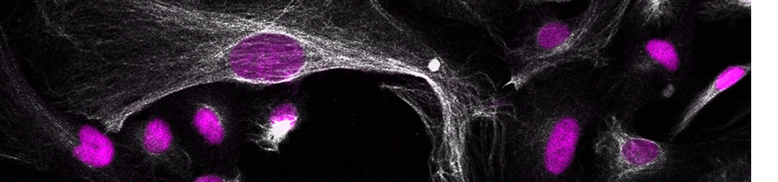
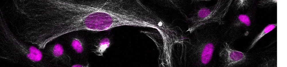
Pubmed AbstractDuring embryonic development, excitatory projection neurons migrate in the cerebral cortex giving rise to organised layers. Periventricular heterotopia (PH) is a group of aetiologically heterogeneous disorders in which a subpopulation of newborn projection neurons fails to initiate their radial migration to the cortex, ultimately resulting in bands or nodules of grey matter lining the lateral ventricles. Although a number of genes have been implicated in its cause, currently they only satisfactorily explain the pathogenesis of the condition for 50% of patients. Novel gene discovery is complicated by the extreme genetic heterogeneity recently described to underlie its cause. Here, we study the neurodevelopmental role of endothelin-converting enzyme-2 (ECE2) for which two biallelic variants have been identified in two separate patients with PH. Our results show that manipulation of ECE2 levels in human cerebral organoids and in the developing mouse cortex leads to ectopic localisation of neural progenitors and neurons. We uncover the role of ECE2 in neurogenesis, and mechanistically, we identify its involvement in the generation and secretion of extracellular matrix proteins in addition to cytoskeleton and adhesion.
Di Matteo F*, Pipicelli F*, Kyrousi C, Tovecci I, Penna E, Crispino M, Chambery A, Russo R, Ayo‐Martin AC, Giordano M, et al. Cystatin B is essential for proliferation and interneuron migration in individuals with EPM 1 epilepsy. * Equal contributing authors. EMBO molecular medicine [Internet]. 2020;12(6):e11419.
Pubmed AbstractProgressive myoclonus epilepsy (PME) of Unverricht-Lundborg type (EPM1) is an autosomal recessive neurodegenerative disorder with the highest incidence of PME worldwide. Mutations in the gene encoding cystatin B (CSTB) are the primary genetic cause of EPM1. Here, we investigate the role of CSTB during neurogenesis in vivo in the developing mouse brain and in vitro in human cerebral organoids (hCOs) derived from EPM1 patients. We find that CSTB (but not one of its pathological variants) is secreted into the mouse cerebral spinal fluid and the conditioned media from hCOs. In embryonic mouse brain, we find that functional CSTB influences progenitors' proliferation and modulates neuronal distribution by attracting interneurons to the site of secretion via cell-non-autonomous mechanisms. Similarly, in patient-derived hCOs, low levels of functional CSTB result in an alteration of progenitor's proliferation, premature differentiation, and changes in interneurons migration. Secretion and extracellular matrix organization are the biological processes particularly affected as suggested by a proteomic analysis in patients' hCOs. Overall, our study sheds new light on the cellular mechanisms underlying the development of EPM1.
Tsaridou S, Skamnelou M, Iliadou M, Lokka G, Parlapani E, Mougkogianni M, Danalatos R-I, Kanellou A, Chlorogiannis D-D, Kyrousi C, et al. Three-dimensional models for studying neurodegenerative and neurodevelopmental diseases. GeNeDis 2018: Genetics and Neurodegeneration [Internet]. 2020:35-41.
Pubmed AbstractHuman brain possesses a unique anatomy and physiology. For centuries, methodological barriers and ethical challenges in accessing human brain tissues have restricted researchers into using 2-D cell culture systems and model organisms as a tool for investigating the mechanisms underlying neurological disorders in humans. However, our understanding regarding the human brain development and diseases has been recently extended due to the generation of 3D brain organoids, grown from human stem cells or induced pluripotent stem cells (iPSCs). This system evolved into an attractive model of brain diseases as it recapitulates to a great extend the cellular organization and the microenvironment of a human brain. This chapter focuses on the application of brain organoids in modelling several neurodevelopmental and neurodegenerative diseases.
Drechsel J, Kyrousi C, Cappello S, Sieber SA.
Tranylcypromine specificity for monoamine oxidase is limited by promiscuous protein labelling and lysosomal trapping. RSC Chemical Biology [Internet]. 2020;1(4):209-213.
Pubmed AbstractMonoamine oxidases MAOA and MAOB catalyze important cellular functions such as the deamination of neurotransmitters. Correspondingly, MAO inhibitors are used for the treatment of severe neuropsychiatric disorders such as depression. A commonly prescribed drug against refractory depression is tranylcypromine, however, the side effects are poorly understood. In order to decipher putative off-targets, we synthesized two tranylcypromine probes equipped with either an alkyne moiety or an alkyne-diazirine minimal photocrosslinker for in situ proteome profiling. Surprisingly, LC-MS/MS analysis revealed low enrichment of MAOA and relatively promiscuous labeling of proteins. Photoprobe labeling paired with fluorescent imaging studies revealed lysosomal trapping which could be largely reverted by the addition of lysosomotropic drugs.
Ayo-Martin AC, Kyrousi C, Di Giaimo R, Cappello S.
GNG5 controls the number of apical and basal progenitors and alters neuronal migration during cortical development. Frontiers in Molecular Biosciences [Internet]. 2020;7:578137.
Pubmed AbstractCortical development is a very complex process in which any temporal or spatial alterations can give rise to a wide range of cortical malformations. Among those malformations, periventricular heterotopia (PH) is characterized by clusters of neurons that do not migrate to the correct place. Cerebral organoids derived from patients with mutations in DCHS1 and FAT4, which have been associated with PH, exhibit higher levels of GNG5 expression in a patient-specific cluster of neurons. Here we investigate the role of GNG5 during the development of the cerebral cortex in mice and human cerebral organoids. GNG5, highly expressed in progenitors and downregulated in neurons, is critical for controlling the number of apical and basal progenitors and neuronal migration. Moreover, forced expression of GNG5 recapitulates some of the alterations observed upon downregulation of Dchs1 and Fat4 in mice and human cerebral organoids derived from DCHS1 and FAT4 patients, suggesting a critical role of GNG5 in cortical development.